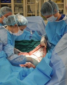
CALEC surgery represents a groundbreaking advancement in the field of eye surgery, offering renewed hope for patients with previously untreatable corneal damage. Developed by Ula Jurkunas at Mass Eye and Ear, this innovative procedure utilizes stem cell therapy to restore the surface of the cornea by cultivating limbal epithelial cells from a healthy eye. In a clinical trial, CALEC surgery demonstrated over 90 percent efficacy in regenerating corneal tissues, significantly improving the vision of participants suffering from limbal stem cell deficiency. As researchers pursue this exciting intervention, the potential for CALEC to revolutionize corneal repair and vision restoration continues to grow. This procedure not only promises a new frontier in treating ocular injuries but also highlights the transformative role of stem cell technology in medicine.
Known as cultivated autologous limbal epithelial cell (CALEC) transplantation, this surgical method harnesses the power of stem cell therapy to facilitate corneal restoration. By extracting healthy limbal epithelial cells from an unaffected eye, doctors can manufacture a viable graft that promotes healing in damaged corneas. This innovative approach not only addresses serious injuries that lead to vision impairment but also sets the stage for future advancements in ocular therapies. As we explore safer and more effective techniques, the restoration of sight through corneal repair becomes an attainable goal for more patients than ever before. The implications of such early successes in this field open doors to expanded applications of stem cell treatments in ophthalmology.
Understanding CALEC Surgery
CALEC surgery, or Cultivated Autologous Limbal Epithelial Cell therapy, represents a significant advancement in the field of ocular medicine. This innovative procedure involves harvesting healthy limbal epithelial cells from a patient’s unaffected eye, then cultivating these cells to create a graft that can be transplanted into the damaged eye. The meticulously engineered graft works to restore the corneal surface, effectively addressing conditions that previously resulted in permanent vision loss. This technique not only highlights the potential of regenerative medicine but underscores the importance of personalized treatment in ophthalmology.
The success of CALEC surgery stems from the ability to harness the body’s own cells to facilitate healing, offering a promising alternative for patients suffering from limbal stem cell deficiency. Through the careful observation of patients over an 18-month period in a clinical trial, researchers demonstrated that CALEC surgery achieved significant restoration of the cornea. The results show not just physical improvements, but a profound positive impact on the quality of life for individuals with previously untreatable eye injuries.
The Role of Stem Cell Therapy in Eye Surgery
Stem cell therapy is revolutionizing eye surgery and treatment options for patients with corneal damage. The ability to utilize stem cells, particularly cultivated from a healthy eye, is paving the way for more effective interventions that restore vision. In the context of CALEC surgery, stem cell therapy serves as a powerful tool that facilitates the regeneration of the corneal surface by replenishing lost limbal epithelial cells. This innovative approach not only repairs physical damage but also addresses the underlying cellular deficiencies that contribute to visual impairment.
In clinical trials, stem cell therapy has demonstrated impressive efficacy, with CALEC treatment yielding a high success rate in restoring corneal integrity. Researchers are excited about the potential implications of these findings, as the effective restoration of the cornea can lead to dramatic improvements in visual acuity and overall patient satisfaction. With ongoing studies exploring the full capabilities of stem cell application in eye surgery, the future looks bright for patients suffering from debilitating eye conditions.
The clinical application of stem cell therapy extends beyond just corneal repair; it suggests a broader potential for vision restoration across various eye conditions. As the medical community increasingly embraces stem cell innovations, the possibilities for treating or even reversing certain types of blindness are becoming more attainable.
The Impact of Corneal Repair Techniques
Corneal repair techniques, particularly through advanced methods like CALEC surgery, have brought new hope to those who once faced chronic pain and visual difficulties. By restoring the corneal surface, these techniques improve both the visual and functional aspects of the eye for patients affected by serious injuries or conditions like chemical burns and infections. The technology behind graft manufacturing has advanced significantly, helping researchers meet strict safety criteria for human trials.
Moreover, successful corneal repair using CALEC not only alleviates physical discomfort associated with corneal damage but also enhances patients’ quality of life, allowing them to regain independence and improve their day-to-day activities. As researchers continue to refine corneal repair methods and begin larger-scale trials, the expectation is that many more patients will benefit from these groundbreaking advancements in eye surgery.
Limbal Epithelial Cells and Their Function
Limbal epithelial cells play a crucial role in maintaining the health and integrity of the cornea. Positioned at the border between the cornea and the sclera, these cells are essential for protecting the eye’s surface and fostering a regenerative environment. When these cells are lost due to injury or disease, the eye cannot heal itself naturally, leading to persistent damage and visual impairment.
The cultivation of limbal epithelial cells for use in CALEC surgery shines a light on their significance within ocular health. By utilizing the patient’s own cells to restore the corneal surface, this innovative approach not only addresses immediate visual rehabilitation but also taps into the body’s natural healing processes, enhancing the potential for successful outcomes in patients with previously untreatable conditions.
Vision Restoration: A New Era with CALEC
The potential for vision restoration through cutting-edge procedures like CALEC surgery marks a new era in ophthalmology. By making use of patient-specific stem cells, this approach allows clinicians to provide tailored treatment options that can lead to substantial improvement in visual acuity and overall eye health. The findings from recent clinical trials demonstrate how effectively CALEC can restore corneal surfaces and provide hope for those with severe corneal deficiencies.
As the data supports an impressive success rate in vision restoration, the implications of CALEC surgery extend beyond personal recovery; they signify a monumental shift in how we approach eye care and treatment. Researchers advocate for continued exploration and the expansion of this treatment modality, seeing the potential for CALEC not only to restore vision but to enhance the quality of life for countless individuals facing debilitating ocular conditions.
The Future of Eye Surgery Innovations
Looking towards the future, innovations in eye surgery, particularly those incorporating stem cell therapy like CALEC surgery, promise to redefine treatment standards for ocular diseases. As the technology and methodologies continue to evolve, it is expected that more patients will gain access to advanced surgical techniques that were once limited to experimental phases. Potential allogeneic approaches could mean that even patients with bilateral corneal damage may soon find relief through similar regenerative strategies.
With continuous investments in research and development, the evolution of eye surgery is anticipated to yield greater successes in restoring vision and managing eye health. The prospects of combining stem cell therapy with new biotechnological advancements present an exciting frontier in ophthalmology, symbolizing hope for effective interventions for various visual impairments.
Clinical Trials and Regulatory Pathways for CALEC
The clinical trials surrounding CALEC surgery represent a foundation for understanding the safety and efficacy of stem cell therapies in ophthalmology. Sponsored by the National Eye Institute, these trials not only assess the therapeutic benefits but also pave the way for future regulatory considerations and potential FDA approvals. Following the positive outcomes observed in initial studies, there is growing anticipation around the establishment of standardized protocols that ensure safe implementation of such innovative procedures.
As researchers and clinicians work together to gather more data, the hope is to further streamline the process for bringing CALEC and similar therapies to the mainstream. The pathway for regulatory approval encompasses various stages, requiring extensive documentation of safety and effectiveness. It is a dynamic process that emphasizes transparency and thoroughness, ensuring patient well-being while promoting advancements in therapeutic options.
Challenges and Limitations of CALEC Surgery
While CALEC surgery has shown remarkable potential, there are inherent challenges and limitations to consider. Notably, one significant requirement is that patients must have an unaffected eye from which to harvest limbal epithelial cells. This limitation poses difficulties for individuals suffering from bilateral damage and emphasizes the need for alternative strategies, such as the exploration of allogeneic stem cell sources.
Moreover, as with any experimental procedure, careful monitoring and ongoing research are crucial to ensure the longevity and success of the treatment. Understanding the long-term implications and potential risks associated with CALEC surgery will help in refining the approach and optimizing patient outcomes, allowing for the development of improved methodologies in corneal repair.
The Importance of Multidisciplinary Collaboration
The journey of developing CALEC surgery highlights the importance of multidisciplinary collaboration in advancing medical treatments. The partnership between various institutions, including Mass Eye and Ear and Dana-Farber, has been pivotal in blending the expertise of researchers, clinicians, and manufacturing specialists to create a cohesive strategy for stem cell treatment. Such collaborations drive the innovation needed to push the boundaries of what is currently achievable in eye surgery.
Moreover, fostering a culture of teamwork amongst various specialties accelerates the pace of discovery, enabling them to tackle complex challenges in eye health. As evidence continues to mount in support of CALEC surgery, collaborative efforts will be essential in expanding treatment options and ensuring equitable access to these therapies for diverse patient populations.
Frequently Asked Questions
What is CALEC surgery and how does it help in eye surgery?
CALEC surgery, or cultivated autologous limbal epithelial cell transplantation, is an innovative eye surgery that utilizes stem cells from a healthy eye to restore the damaged cornea in patients with limbal stem cell deficiency. This procedure has shown over 90% effectiveness in regenerating the corneal surface, offering new hope for those who previously faced untreatable corneal injuries.
How does CALEC surgery utilize stem cell therapy for vision restoration?
CALEC surgery employs stem cell therapy by harvesting limbal epithelial cells from a patient’s healthy eye, expanding them, and transplanting the resulting graft into the affected eye. This approach not only facilitates vision restoration but also addresses corneal repair in cases where traditional methods may not be viable.
Who is eligible for CALEC surgery and what conditions does it treat?
CALEC surgery is primarily for patients with corneal injuries resulting in limbal stem cell deficiency, typically caused by chemical burns, infections, or trauma. Eligibility requires that patients have one unaffected eye from which to harvest the necessary cells.
What are the results of clinical trials involving CALEC surgery?
Clinical trials for CALEC surgery have reported a 93% success rate in restoring the cornea’s surface after 12 months, with significant improvements in visual acuity for many participants. The high safety profile, with minimal adverse events, further supports its potential as a breakthrough in eye surgery.
Is CALEC surgery widely available, and what are the future prospects?
Currently, CALEC surgery remains experimental and is not widely available in hospitals, including Mass Eye and Ear. Future studies aim to include more patients and facilitate FDA approval, potentially expanding access to this promising treatment for eye damage.
What makes CALEC surgery a unique approach to corneal repair?
CALEC surgery is unique because it uses the patient’s own stem cells to create a graft, minimizing rejection risks and providing a personalized treatment option for those with severe corneal damage that was once deemed untreatable.
What are limbal epithelial cells and their role in CALEC surgery?
Limbal epithelial cells are essential for maintaining the cornea’s health and clarity. In CALEC surgery, these cells are harvested from a healthy eye, cultivated, and then transplanted to repair the damaged cornea, thereby restoring its surface and improving vision.
What should patients expect during the CALEC surgery process?
Patients undergoing CALEC surgery can expect an initial biopsy of the healthy eye to obtain limbal epithelial cells, followed by a two to three-week waiting period for cell cultivation. The surgical procedure then involves transplanting the cell graft into the affected eye, typically performed under local anesthesia with a focus on safety and effectiveness.
What are the limitations of CALEC surgery?
One primary limitation of CALEC surgery is that it can currently only be performed on patients with one involved eye, as it requires a healthy eye to extract the necessary cells. Future developments may aim to use donor cells to broaden the eligibility criteria for this treatment.
| Key Aspect | Details |
|---|---|
| Procedure Name | Cultivated Autologous Limbal Epithelial Cells (CALEC) |
| First Surgery Location | Mass Eye and Ear |
| Principal Investigator | Ula Jurkunas |
| Success Rate (after 18 months) | 92% overall success rate, with 93% complete restoration |
| Eligibility for Treatment | Patients with one healthy eye for biopsy required |
| Outcome | Restored cornea in 50% of patients at 3 months, increasing over time |
| Safety Profile | No serious adverse events; one minor infection reported |
| Future Prospects | Hopes to establish allogeneic manufacturing for broader use |
Summary
CALEC surgery represents a groundbreaking advancement in the treatment of corneal injuries deemed untreatable. This innovative technique utilizing stem cells from a healthy eye showcases tremendous promise, with a notable 92% success rate in restoring corneal surfaces over an 18-month period. The research spearheaded by Ula Jurkunas at Mass Eye and Ear has opened new avenues for patients suffering from vision-impairing corneal damage. As ongoing clinical trials continue to expand on these findings, the ultimate goal is to make CALEC surgery accessible to a broader patient demographic, potentially transforming the landscape of ocular rehabilitation.
